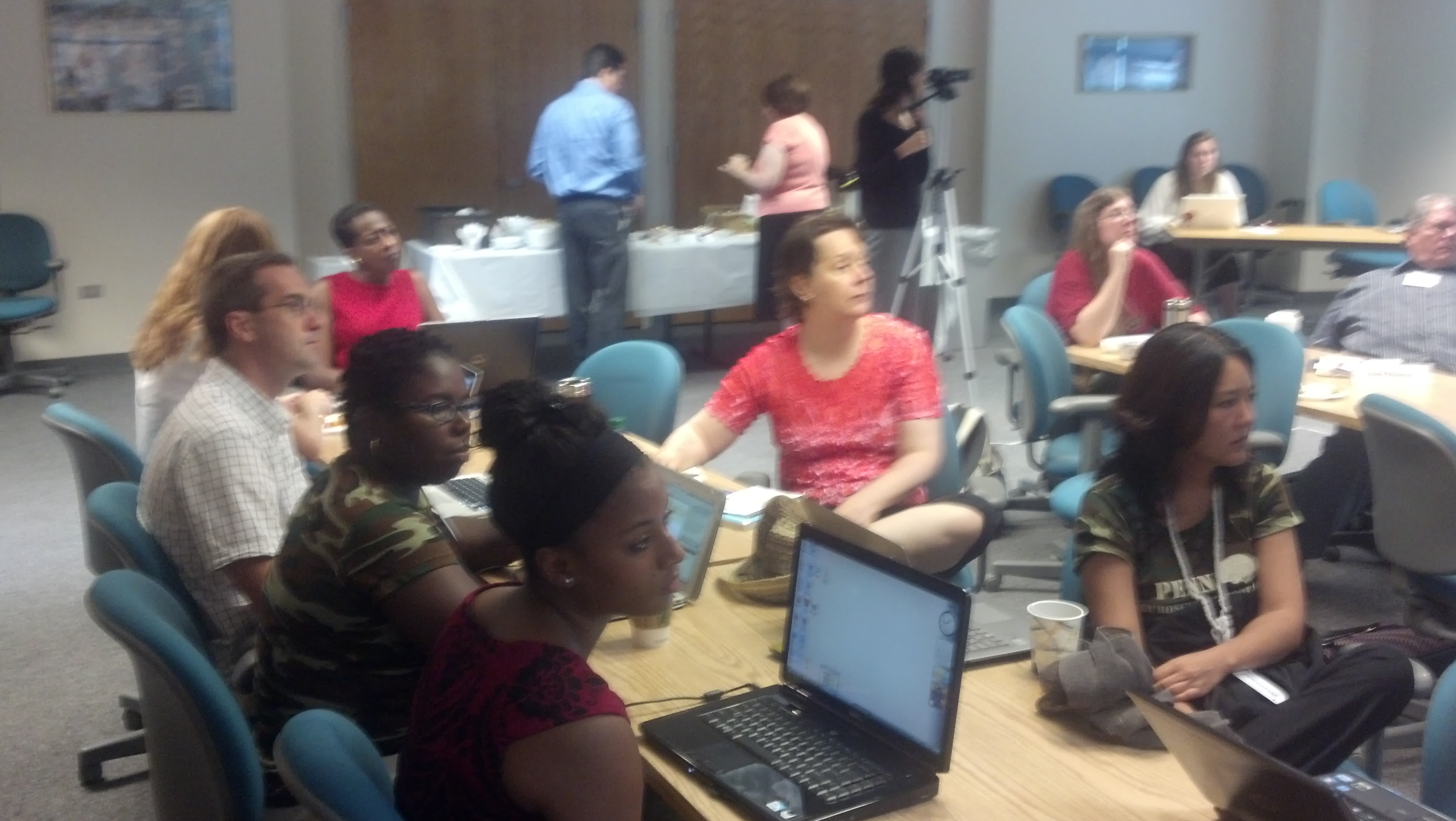
A long but beneficial day studying emotion and the brain. Dr. Joseph Kable provided three lectures that started with the basics of neural correlates to emotion (regions involved), the nature of motivation including specific issues involved in addiction, the place of decision making in emotion, and the role of empathy both with social interactions and moral decision making (which include both judgement and empathy). There were just too many insights for summary, and some of the material had to be condensed with an extensive discussion over methodology (n0t surprising with a group this size). Still, a lot of important material introduced. Rather than cover everything I am just going to touch on a few highlights that might intersect with popular ideas.
One preliminary, Kable defined emotion as a “discrete response to an external or internal event involving a synchronized:
- expression
- bodily response
- subjective experience or feeling
- evaluation and appraisal
- action tendencies”
Kable notes psychologists prefer to use terms like intensity and valence (toward positive or negative) rather than terms like “joy” or “love” since the more technical terms provide better control for the sake of comparison (so joy becomes an intense positive emotion and fear an intense negative emotion).
One surprise to me is that the historically used idea of the limbic system is losing favor in neuroscience with greater interest in researching the Dopamine/Reward correlation and the Amygdala/Fear correlation.
One caution is that neuroscience is always using comparative frameworks so one traditional study involved standard behavior reward (Pavlov).
 Dopamine seems to be active both when an animal receives an unexpected reward but later appears when the animal “anticipates” the reward (we are talking milliseconds) and drops rapidly if the reward does not appear. However the study tested the difference between receiving or not receiving the reward (anticipatory set not the issue). Dopamine is not encoding the presence of the reward but encoding a reward prediction error (what you got versus what you expected to get). However, the overall study proves helpful since it helps us understand learning through “trial and error” based on the number of tstwe do not get it correct). This simple study opens up a neurochemical basis for the most basic form of learning.
Similarly studies on fear (see the work of Joesph LeDoux) provide a basic learning theory. Think of the fact animals “freeze” at the sight of a dangerous snake or when a sound emits a dangerous shock. 
Amygdala is one place where all information occurs in brain to address this phenomenon: lateral nucleus has ability to gather both sound and shock while central nucleus to mediate freezing, blood pressure, and hormones. Also fear studies contribute to the idea “cells that fire together also wire together.” The concept is called long term potentiation (LTP) where continued synaptic firing between two neurons (axon to dendrite) “strengthen” the connection between the two neurons (via amplification: the strength of the response is greater to the firing). This provides a synaptic basis for learning as well (LeDoux says we are “synaptic selves”).
Later in the day we moved beyond these basic categories to discuss judgment and emotion. It is important that controlling emotion is not just the Prefrontal Cortext “shutting down” the seat of emotions (amygdala) but actually activates the subcortical striatum
 (which has densest number of dompamanergic receptors) so that the PFC (seat of executive function) actually “biases” flow of activation in other areas of the brain based on the representation of what the rules are in the PFC. So PFC is not directly controlling but modulating stimulus/response. A question rose whether to call the brain’s executive function a Modulator? Mediator? perhaps Moderator is best term. Raises question whether a better metaphor for brain activity might be harmonics rather than hydraulics… something to reflect on later.
When engaged in empathy or social decision making Kable noted we have to begin with a theory of the mind (great example is the following movie Heider-Simmel Demonstration where we ascribe intention to actions (even if just objects).
 Empathy seems to involve the same areas of the brain where we directly feel pain and pleasure (so seeing someone in pain lights up the same parts of the brain as experiencing pain, but also just telling someone of a painful event!)
 When it comes to social interaction and moral decisions, we seem to employ both decision making processes (theory of mind area) and empathetic pathways in many “personal” decisions. Need to end this post. Much more to say but a good day to “digest” in the coming weeks.